Introduction to Medium to Deep Chemical Peels
Acne can be persistent and difficult to manage, especially when it leads to scarring, inflammation, and deep breakouts. Medium to deep chemical peels offer an advanced treatment option that targets severe acne, clogged pores, and post-inflammatory hyperpigmentation. These peels penetrate deeper into the skin layers, effectively removing damaged skin cells, regulating oil production, and stimulating skin renewal for a clearer, smoother complexion. Medium to deep peels use stronger exfoliating agents such as trichloroacetic acid (TCA), phenol, or high-strength salicylic acid to reach the mid to lower dermis, making them effective for stubborn acne, deep scars, and long-term skin rejuvenation.
A skin biopsy is the definitive diagnostic tool in dermatology — it provides histopathological evidence that clinical and dermoscopic examination alone cannot always deliver. At Claire Derma, we perform biopsies when a skin lesion's appearance raises diagnostic uncertainty, when a rash fails to respond to standard treatment, or when monitoring detects changes in a previously stable mole.
How Medium to Deep Chemical Peels Work
These peels work by deeply penetrating the skin to:
Determining whether a biopsy is indicated — and selecting the correct biopsy type — is a clinical decision that depends on the suspected diagnosis. At Claire Derma, our dermatologists consider the lesion's size, depth, anatomical location, and the differential diagnoses being considered.
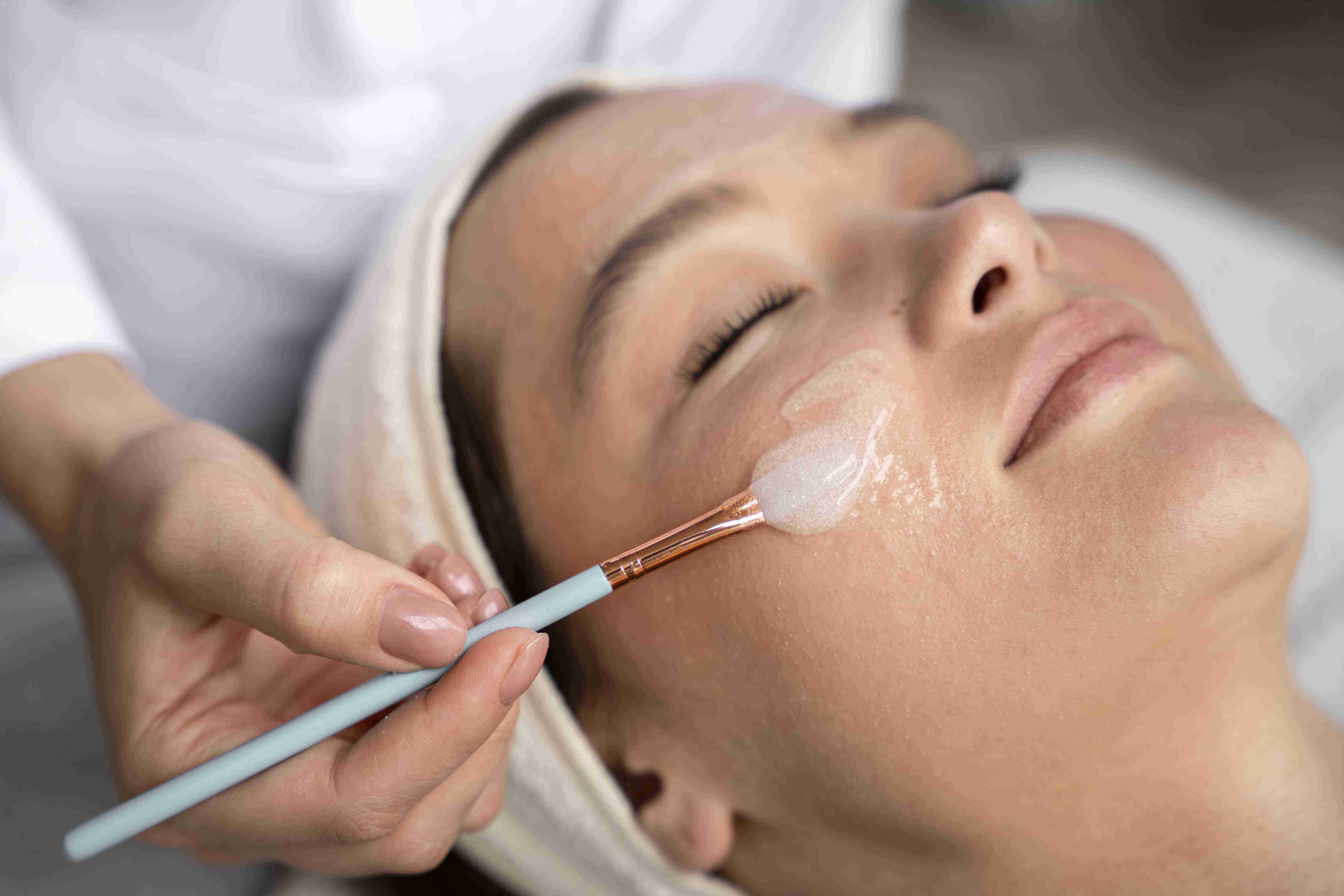
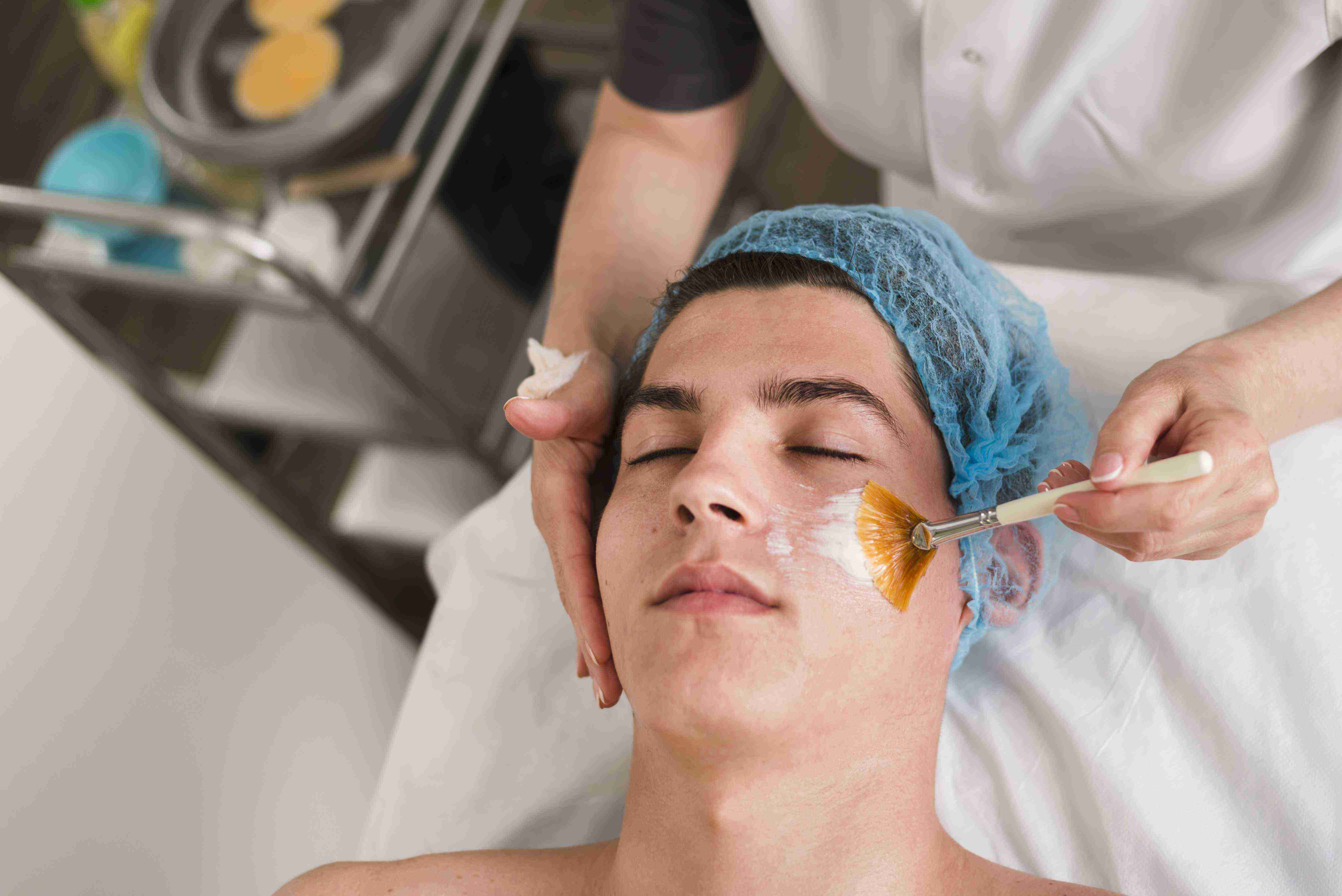
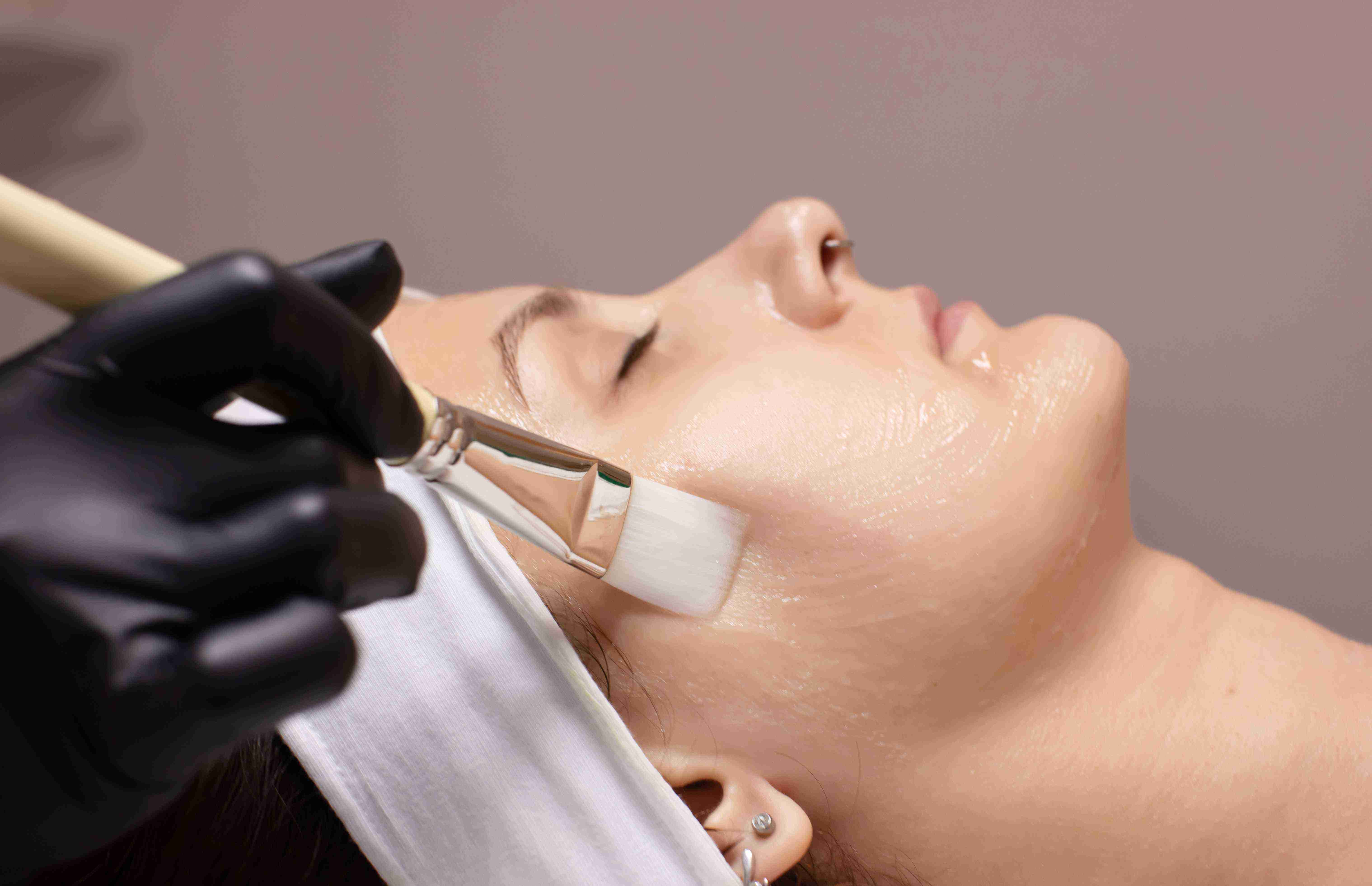
Treating Acne with Medium to Deep Chemical Peels
Biopsy technique at Claire Derma is precise and tailored to the diagnostic question. Punch biopsies use a circular blade — typically three to six millimetres — that cuts through the epidermis and dermis to the subcutaneous fat in a single motion. The cylindrical core is lifted with forceps and the wound is closed with one or two sutures.
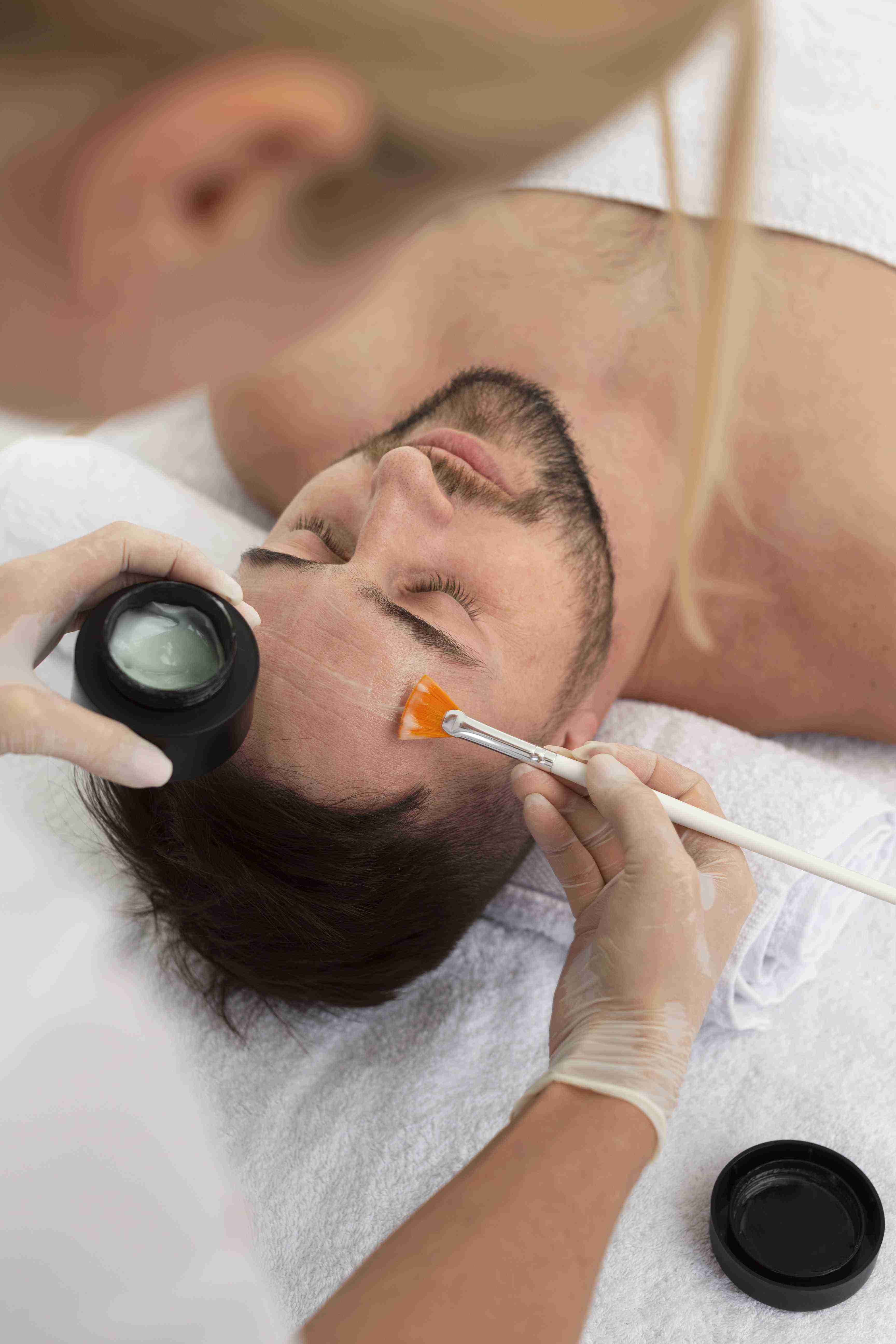
Benefits of Medium to Deep Peels for Acne
The diagnostic value of a properly performed skin biopsy cannot be replicated by any non-invasive method currently available. While dermoscopy, confocal microscopy, and AI-assisted imaging tools have improved clinical accuracy, they provide pattern-based probability rather than tissue-level certainty. A biopsy examined by a dermatopathologist reveals the cellular composition, architectural organisation, and depth of involvement that define the diagnosis.
Ideal Candidates for the Treatment
This treatment is ideal for individuals who:
Biopsy site care at Claire Derma is designed to be simple and easy to follow. Punch and excisional biopsy sites are closed with sutures and covered with a small adhesive dressing. Keep the area dry for the first 24 hours, then clean gently with water and reapply a thin layer of petroleum jelly with a fresh dressing daily. Avoid submerging the wound in baths, pools, or hot tubs until sutures are removed.
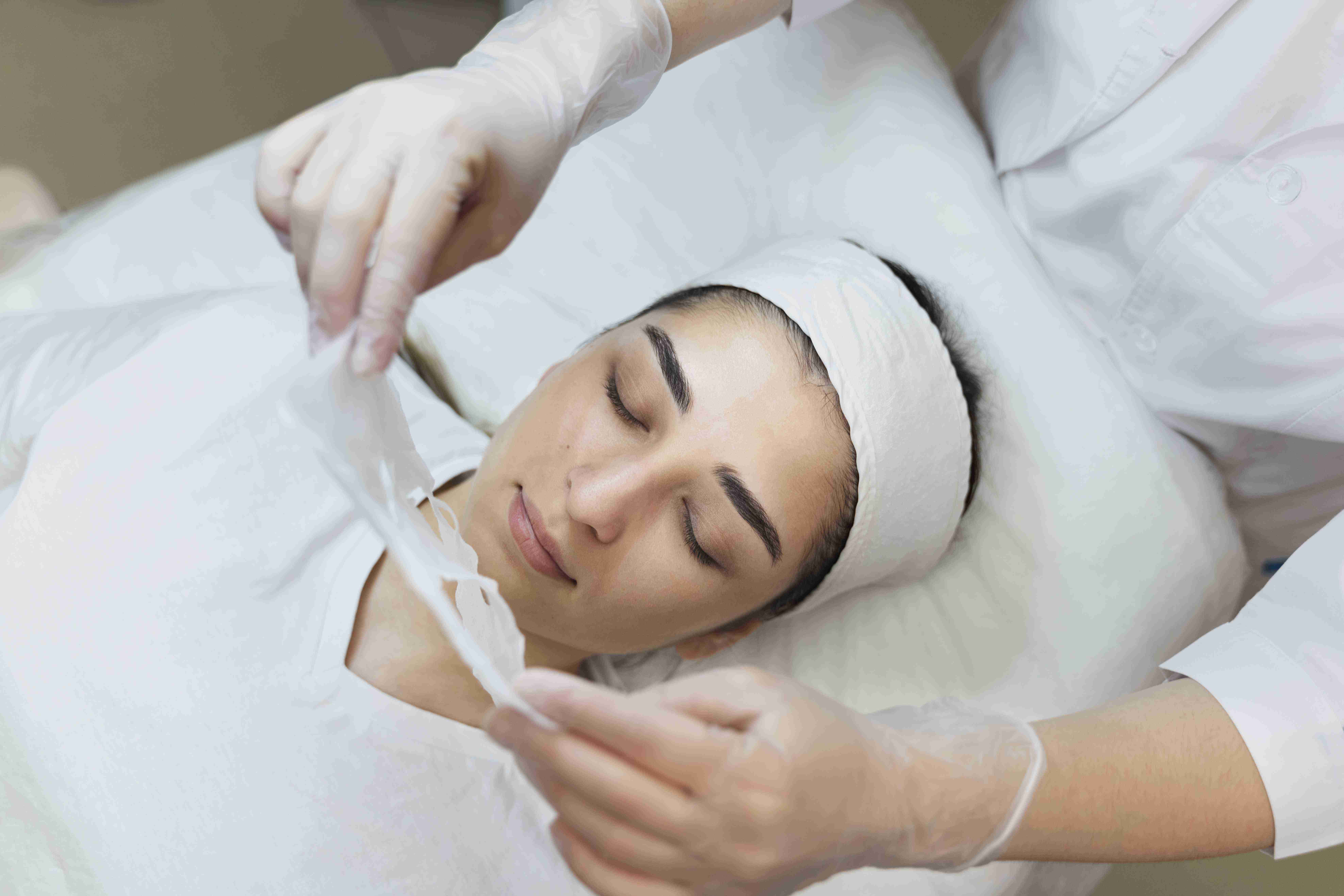
The Treatment Process
01
Consultation & Skin Analysis
The dermatologist assesses acne severity and selects the best peel formulation. Your dermatologist examines the lesion or rash, reviews your medical history and any prior treatments, and explains why a biopsy is recommended. The biopsy type — punch, shave, or excisional — is selected based on the suspected diagnosis and lesion characteristics.
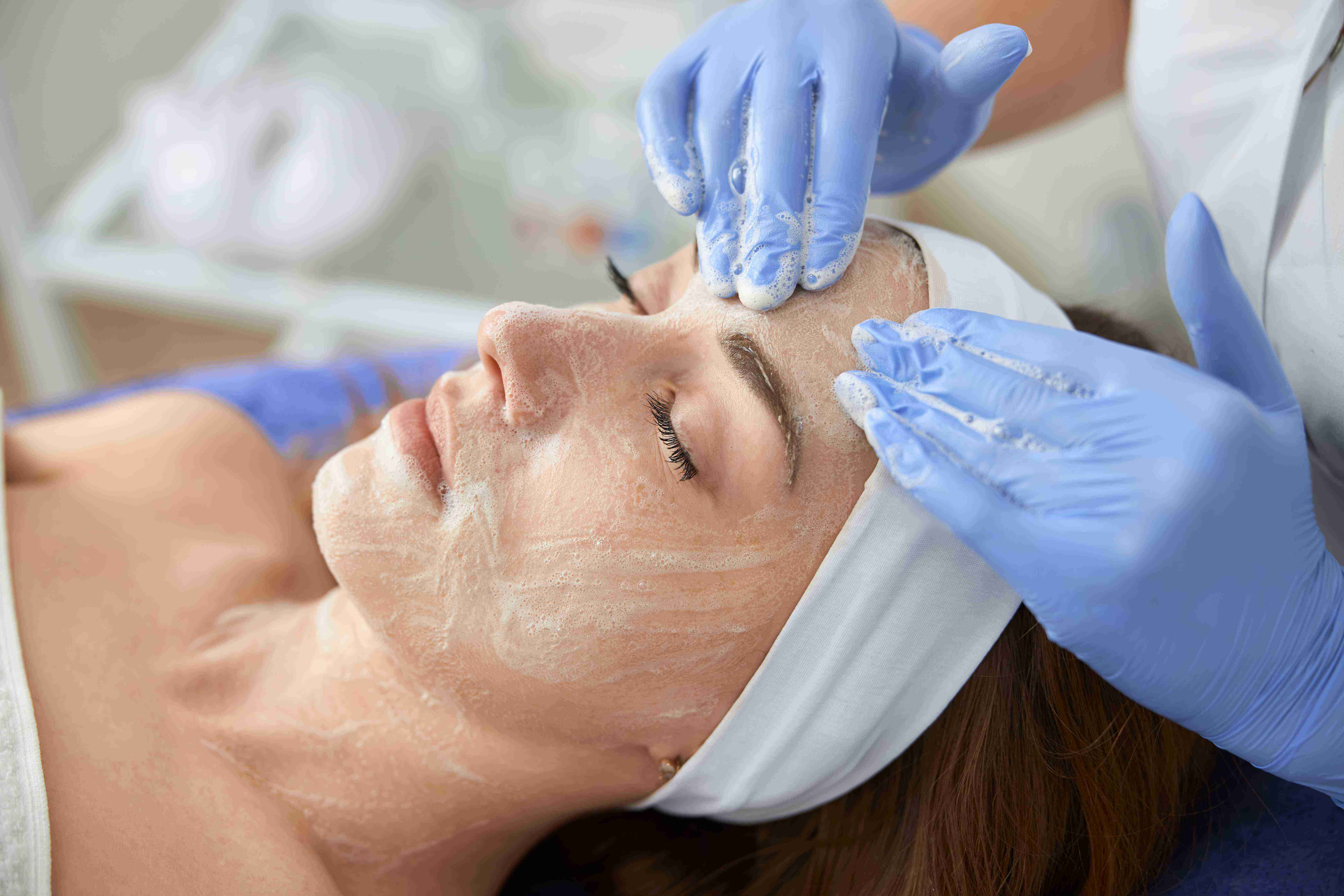
02
Preparation
The skin is cleansed, and protective measures are taken for sensitive areas. The biopsy site is cleaned with chlorhexidine antiseptic. Local anaesthesia — typically one percent lidocaine with adrenaline — is injected using a fine-gauge needle. The injection produces a brief sting lasting two to three seconds.
03
Peel Application
A customized medium or deep peel solution is applied for controlled penetration. The biopsy is performed using the selected technique. Punch biopsies take under one minute. Shave biopsies take thirty seconds to one minute. Excisional biopsies take ten to fifteen minutes including suturing.
04
Neutralization & Post-Treatment Care
The peel is neutralized, and a soothing recovery serum is applied. The biopsy site is dressed with a simple adhesive dressing. Written aftercare instructions are provided. Suture removal is scheduled at the appropriate interval. Histopathology results are communicated within five to seven working days.
Expected Results & Recovery
Visible improvements after one session, with further refinement over multiple treatments.
Fewer blemishes, reduced scarring, and an even complexion.
Expect peeling and skin sensitivity for 5–7 days.
With proper maintenance, results can last several months.
Got Questions?We've Got Answers
Find answers to the most common questions about our treatments, procedures, and recovery process. If you can't find what you're looking for, our support team is always here to help.
Typically, 3–6 sessions, spaced 4–6 weeks apart, yield the best results.
Patients may feel a warm or stinging sensation, but discomfort is manageable.
Peeling and flaking can last between 5–10 days, depending on peel strength.
It significantly reduces acne and breakouts, but maintenance treatments and a proper skincare routine are essential.
Sun exposure must be strictly avoided for at least a week, and broad-spectrum sunscreen is mandatory.
The local anaesthetic injection produces a brief sting lasting two to three seconds — after that, the biopsy site is completely numb and you will not feel the procedure. Patients often describe the injection discomfort as similar to a mild pinch. At Claire Derma, we use fine-gauge needles and buffered lidocaine to minimise this initial sensation.
Standard histopathology results are available within five to seven working days. Cases that require special stains, immunohistochemistry, or expert second opinion may take up to ten working days. At Claire Derma, we contact you as soon as results are available — either at your suture removal appointment or by phone if results arrive sooner.
All biopsies leave some mark, but the size is proportional to the technique used. A three-millimetre punch biopsy produces a tiny scar the size of a pin dot. Shave biopsies leave a flat pink mark that fades over several weeks. Excisional biopsies produce a thin linear scar similar to any minor surgical wound.
If the histopathology report identifies a skin cancer, precancerous condition, or systemic disease, our dermatologist will contact you promptly to discuss the findings and plan the next steps. For skin cancers, this may involve wider excision with clear margins or referral to a specialist surgeon for Mohs micrographic surgery. For inflammatory conditions, appropriate systemic or topical therapy is initiated.